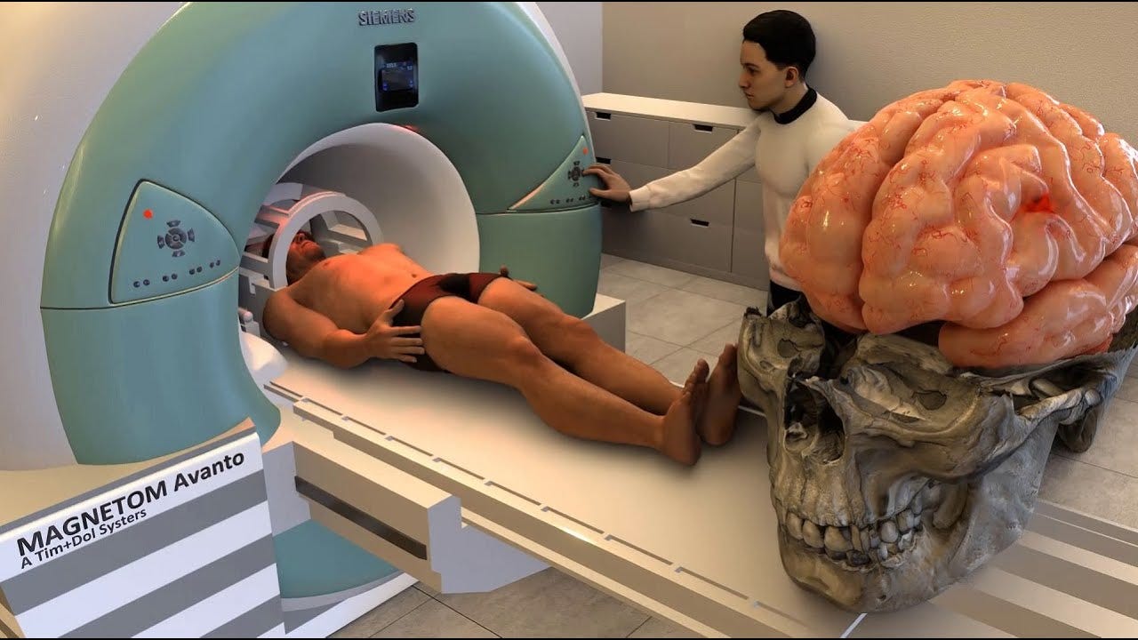
How Exactly Does An MRI Work?
Written by Umme Rokshana
Magnetic Resonance Imaging (MRI) is a powerful medical tool that allows doctors to see inside the human body without using invasive procedures or harmful radiation. Sure, it’s one of the most needed technologies out there, but how exactly does it work?
Nuclear magnetic resonance: Human bodies are made up of a large amount of water, which contains hydrogen atoms. These hydrogen atoms have nuclei that behave like tiny magnets because of the presence of protons (positively charged particles). When you lie inside an MRI scanner, a strong magnetic field—usually thousands of times stronger than the Earth's magnetic field—aligns these hydrogen nuclei in your body.
Next, the MRI machine sends out a pulse of radio waves that momentarily disrupts this hydrogen alignment. When the pulse stops, the hydrogen nuclei gradually return to their original alignment, and in doing so, they release energy in the form of radio signals. These signals are detected by the scanner and sent to a computer, which processes them to create highly specific/detailed images of the body's internal structures.
What makes MRI especially useful is its ability to differentiate between various types of tissues. For example, healthy tissues and abnormal growths like tumors have different water content and therefore emit different signals, allowing radiologists to identify and diagnose conditions that may not be visible on X-rays or CT scans.
MRI technology continues to evolve, offering clearer images and faster scan times, making it an indispensable tool in modern medicine. Whether it’s for brain imaging, joint analysis, or spotting abnormalities in soft tissues, MRIs provide a non-invasive, highly accurate way to diagnose and monitor various medical conditions.
References
https://www.nibib.nih.gov/science-education/science-topics/magnetic-resonance-imaging-mri
Written by Umme Rokshana from MEDILOQUY


