
Cryo-EM
Written by Christopher Dass

Introduction:
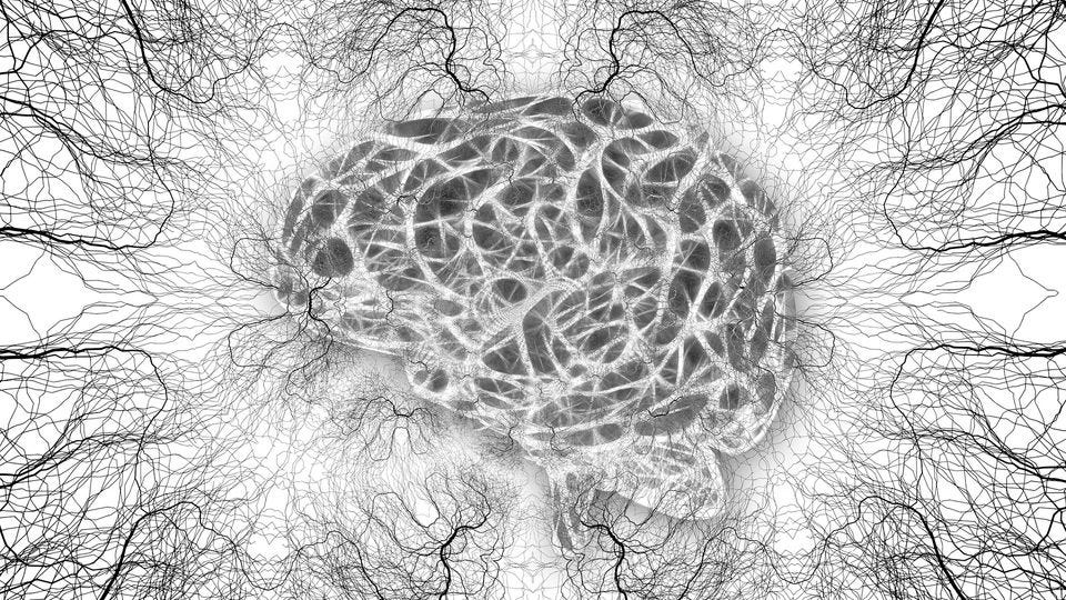
Studying the human brain has always been a challenge for scientists. Cryo-electron microscopy (Cryo-EM) now offers highly detailed images of brain structures at nearly atomic levels, providing unprecedented insights into how the brain works at a molecular level.
Understanding Cryo-EM:
Cryo-EM involves freezing biological samples rapidly to extremely low temperatures and then imaging them using an electron microscope. This method preserves the natural state of samples, allowing scientists to observe intricate structures with high resolution and accuracy, without the distortions seen in traditional preparation methods.
Impact on Brain Research:
Cryo-EM has transformed the study of brain structures by revealing detailed images of synapses, receptors, and other essential components of neural cells. These images help neuroscientists understand how these structures function and interact, advancing our knowledge of brain activity and neurological diseases.
Technological Progress:
Advances in direct electron detectors and advanced image processing algorithms have enhanced Cryo-EM's capabilities. These improvements enable more precise reconstructions of brain structures, enabling scientists to delve deeper into the complexities of the brain.
Neuroscientific Applications:
Cryo-EM is crucial in examining structural changes associated with neurodegenerative diseases like Alzheimer’s and Parkinson’s. By visualizing these changes at the molecular level, researchers can develop targeted therapies that address the underlying causes of these conditions. Additionally, Cryo-EM aids in drug development by revealing how potential treatments interact with brain structures, facilitating the creation of more effective medications.
Future Prospects:
Continued advancements in Cryo-EM technology may lead to even higher resolution imaging, allowing for real-time study of dynamic brain processes. This could revolutionize our understanding of brain function and lead to the development of new treatments for various neurological disorders.
Conclusion:
Cryo-EM is advancing neuroscience by providing detailed views of the brain's molecular structures. It enhances our understanding of brain function and diseases, driving progress in therapy development. As Cryo-EM continues to evolve, it promises new breakthroughs in brain research and medicine.
Sources:
Author links open overlay panelBenoît Zuber a, a, b, Highlights•Cryo-ET imaging of neurons is a versatile system for cell biology in situ.•Structural and spatial localization analysis yields new insights into synaptic transmission.•The synapse provides a rich environment for the development of image process, & AbstractCryo-electron tomography (Cryo-ET) provides unique opportunities to image cellular components at high resolution in their native state and environment. While many different cell types were investigated by cryo-ET. (2022, March 9). Neurons as a model system for cryo-electron tomography. Journal of Structural Biology: X. https://www.sciencedirect.com/science/article/pii/S2590152422000083
Written by Christopher Dass from MEDILOQUY